Dr Michell Ruiz

Dr Michell Ruiz
Cirugía de hombro y del Manguito rotador

jueves, 11 de junio de 2015

Rehabilitación tras reparación artroscópica del manguito / Delayed versus early motion after arthroscopic rotator cuff repair: a meta-analysis

#reparación artroscopica  #early motion

Fuente
Este artículo es originalmente publicado en:
http://www.ncbi.nlm.nih.gov/pubmed/25127908
http://www.jshoulderelbow.org/article/S1058-2746(14)00318-8/abstract
http://www.anatomia-fisioterapia.es/22-systems/musculoskeletal/upper-extremity/shoulder/1185-rehabilitacion-tras-reparacion-artroscopica-del-manguito
De:
Chan K1MacDermid JC2Hoppe DJ1Ayeni OR1Bhandari M1Foote CJ1Athwal GS3.
 2014 Nov;23(11):1631-9. doi: 10.1016/j.jse.2014.05.021. Epub 2014 Aug 13.
Todos los derechos reservados para:
Copyright © 2014 Journal of Shoulder and Elbow Surgery Board of Trustees. Published by Elsevier Inc. All rights reserved.

Abstract

BACKGROUND:

We conducted a meta-analysis of randomized trials to compare delayed vs early motion therapy on function after arthroscopic rotator cuff repair.

METHODS:

We searched 4 electronic databases (Medline, Embase, Cochrane, and Physiotherapy Evidence Database [PEDro]). The methodologic quality of the included studies was assessed, and the relevant data were extracted. Data were pooled for functional outcomes, rotator cuff tear recurrence, and shoulder range of motion. Complications were reported descriptively.

RESULTS:

Three level I and 1 level II randomized trials were eligible and included. Pooled analysis revealed no statistically significant differences in American Shoulder and Elbow Surgeons scores between delayed vs early motion rehabilitation (mean difference [MD], 1.4; 95% confidence interval [CI], -1.8 to 4.7; P = .38, I(2) = 34%). The risk of retears after surgery did not differ statistically between treatment groups (risk ratio, 1.01; 95% CI, 0.63-1.64; P = .95). Early passive motion led to a statistically significant, although clinically unimportant, improvement in forward elevation between groups (MD, -1°; 95% CI, -2° to 0°; P = 0.04, I(2) = 0%). There was no difference in external rotation between treatment groups (MD, 1°; 95% CI, -2° to 4°; P = 0.63, I(2) = 0%). None of the included studies identified any cases of postoperative shoulder stiffness.

CONCLUSIONS:

The current meta-analysis did not identify any significant differences in functional outcomes and relative risks of recurrent tears between delayed and early motion in patients undergoing arthroscopic rotator cuff repairs. A statistically significant difference in forward elevation range of motion was identified; however, this difference is likely clinically unimportant.
Copyright © 2014 Journal of Shoulder and Elbow Surgery Board of Trustees. Published by Elsevier Inc. All rights reserved.

KEYWORDS:

Rotator cuff; motion; rehabilitation; shoulder; therapy
PMID:
 
25127908
 
[PubMed - indexed for MEDLINE]

Rehabilitación tras reparación artroscópica del manguito

Hasta la fecha, no está claro que tipo de rehabilitación tras la reparación del manguito rotador condice a los resultados más beneficiosos – no habiendo llegado aún a un consenso en la duración de la inmovilización. La rigidez post-cirugía puede disminuir cuando la movilización comienza en una fase temprana aunque un período más largo de restricción de movilidad podría facilitar la curación tendón/hueso.
En esta revisión sistemática con meta-análisis, se incluyó 4 RCT comparando la movilidad temprana con la atrasada. Las medidas de resultado fueron la escala ASES, la escala Constant-Murley, tests simples de hombro, el índice Western Ontario Rotator Cuff y el cuestionario DASH. Se incluyó además en el análisis, el número de recaídas con rotura completa y el ROM de hombro (flexión anterior y rotación externa).
No hubo diferencias significativas en las medidas de resultado, riesgo de roturas recurrentes o ROM de hombro en rotación externa en ambos supuestos. La flexión anterior fue significativamente mayor (pero no clínicamente relevante) en aquellos que recibieron movilización precoz. Basándonos en estos resultados se puede concluir que no existieron diferencias sustanciales a favor de uno de los protocolos de rehabilitación.
> De: Chan et al., J Shoulder Elbow Surg 23 (2015) 1631-1639. Todos los derechos reservados: the Journal of Shoulder and Elbow Surgery Board of Trustees. Pincha aquí para acceder al resumen de Pubmed.. Traducido por Francisco Jimeno Serrano.


miércoles, 3 de junio de 2015

Shoulder arthritis - many different types

Fuente
Este artículo es originalmente publicado en:
http://shoulderarthritis.blogspot.mx/2015/06/shoulder-arthritis-many-different-types.html

Shoulder joint replacement - many different types of arthritis

Degenerative Joint Disease

Degenerative joint disease is also known as osteoarthritis, osteoarthrosis or wear and tear arthritis. The pathogenesis of this condition results from the age-related loss of the ability of articular cartilage to sustain itself against seemingly minor mechanical imbalances and years of use. Degenerative joint disease typically affects healthy and active individuals. In our practice, most patients with degenerative glenohumeral degenerative joint disease are of Northern European ancestry in contrast to those of Asian, Southern European, Hispanic and African ancestry. In degenerative joint disease the glenoid cartilage and subchondral bone are progressively lost - either concentrically medially (more characteristic of female shoulders and those with inflammatory arthritis) or eccentrically posteriorly, with the articular cartilage often left intact anteriorly (more characteristic of male shoulders and those with degenerative joint disease or capsulorrhaphy arthropathy). The concern with the eccentric wear pattern is that the diminished articular contact area results in increased force/unit area leading to increased posterior wear which in turn leads to even less contact area. The cartilage of the humeral head is eroded in a Friar Tuck pattern of central baldness, often surrounded by a rim of remaining cartilage and osteophytes


lunes, 1 de junio de 2015

Suprascapular Nerve Palsy

#Suprascapular Nerve Palsy

Fuente
Este artículo es originalmente publicado en:
http://orthopaedicprinciples.com/2015/06/suprascapular-nerve-palsy/?utm_source=feedburner&utm_medium=email&utm_campaign=Feed%3A+orthopaedicprinciples+%28Orthopaedicprinciples.com+%29
De:
Courtesy: Prof Lennard Funk, Wrightington Upper Limb Unit, Wrightington, UK
Todos los derechos reservados para:
Copyright@orthopaedicprinciples.com. All right rerserved.


jueves, 28 de mayo de 2015

Rotator Cuff MRI

Fuente
Este artículo es originalmente publicado en:
Educational video describing MRI imaging of the rotator cuff.

Become a friend on facebook:
http://www.facebook.com/drebraheim

Follow me on twitter:
https://twitter.com/#!/DrEbraheim_UTMC
  • Categoría

  • Licencia

    • Licencia estándar de YouTube


martes, 26 de mayo de 2015

Reverse total shoulder results and complications

Fuente
Este artículo es originalmente publicado en:
http://shoulderarthritis.blogspot.mx/2015/05/reverse-total-shoulder-results-and.html?utm_source=twitterfeed&utm_medium=twitter

De:

Frederick A. Matsen III, M.D.


Reverse total shoulder results and complications

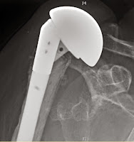
The reverse total shoulder offers a treatment option for a problem that previously had none: the unstable, cuff deficient shoulder. There are now substantial reports of the use of this type of prosthesis to manage a wide range of pathologies, including rotator cuff deficiency without arthritis (see example below in which pseudoparalysis after an attempted tuberosity and cuff repair was treated with a reverse total shoulder)


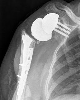
rotator cuff tear arthropathy, rheumatoid arthritis, failed anatomic arthroplasties, arthritis with glenoid bone deficiency, fractures and post traumatic arthritis (as shown in the example below - not that cement was required because the humeral shaft did not allow a secure press fit) 

There are several important failure modes after reverse total shoulder. Infection is one of the more common and is probably related to the fact that many reverse total shoulders are performed as revisions after multiple prior surgeries coupled with the dead space created when the humerus is displaced distally by the procedure. As with anatomic shoulders, Propionibacterium is a frequently cultured organism from failed reverse total shoulders, which can present with loosening in the absence of the usual clinical signs of infection

The infection with Propionibacterium in the x-ray on the left was treated with a single stage exchange to a long stem prosthesis. The patient is currently asymptomatic six years after the revision. 
Instability can result from falls, suboptimal component selection, component malposition, bulky tissues in the posterior shoulder, leverage of the humeral component against the glenoid , or lack of sufficient compressive effect by the deltoid. 
 
Early closed reduction can be successful. Recurrent or chronic instability may require surgical revision. The case below shows a failed anatomic prosthesis for fracture with anterosuperior escape that was treated with a reverse that dislocated recurrently. 
 

The prosthesis was revised to a hemiarthroplasty that was unsatisfactory. Finally a successful revision was accomplished by the removal of posterior scar tissue and revision to a reverse with a 40 mm set of components.

Shown below is another example where stability was restored by changing to a larger diameter of curvature and increasing the thickness of the polyethylene and humeral cup.

The risk of humeral fracture is increased in revision surgery, by falls and when the humeral component fixation results in an abrupt transition between a cemented or press fit diaphyseal stem tip and osteopenic bone distal to the prosthetic tip These fractures deserve a trial at closed management.  
in that surgical revision can be very complex  

Scapular and acromial fractures can result from excessive deltoid tension producing a fatigue fracture or from bone weakened by screw placement. These fractures are preferably treated non-operatively.

Scapular notching is a prominent complication, particularly common in prosthetic designs that medialize the humerus or when the glenoid component is positioned high on the glenoid bone.


The issues with notching are not so much the ‘notch’ in the scapula but the radiographically unseen damage to the polyethylene of the humeral component
 and to glenoid component fixation 
Notching may also be associated with unwanted bone formation that limits the range of motion 
Humeral component failure may result from dissociation of the cup from the stem Glenoid failure may result from glenosphere-baseplate dissociation, glenoid fracture, or failure of fixation While some cases can be reconstructed, others require salvage conversion to a hemiarthroplasty after glenoid component removal; the clinical results of this conversion are generally poor.

Neurologic lesions can result from dissection, retraction or over lengthening of the arm. Finally, there has been some concern about loss of active external rotation with reverse total designs that medialize the tuberosity, prompting consideration of latissimus dorsi transfers; this problem seems less of an issue with those designs that maintain ‘East-West” tensioning of the residual cuff posteriorly.