Dr Michell Ruiz

Dr Michell Ruiz
Cirugía de hombro y del Manguito rotador

jueves, 28 de mayo de 2015

Rotator Cuff MRI

Fuente
Este artículo es originalmente publicado en:
Educational video describing MRI imaging of the rotator cuff.

Become a friend on facebook:
http://www.facebook.com/drebraheim

Follow me on twitter:
https://twitter.com/#!/DrEbraheim_UTMC
  • Categoría

  • Licencia

    • Licencia estándar de YouTube


martes, 26 de mayo de 2015

Reverse total shoulder results and complications

Fuente
Este artículo es originalmente publicado en:
http://shoulderarthritis.blogspot.mx/2015/05/reverse-total-shoulder-results-and.html?utm_source=twitterfeed&utm_medium=twitter

De:

Frederick A. Matsen III, M.D.


Reverse total shoulder results and complications

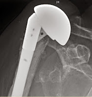
The reverse total shoulder offers a treatment option for a problem that previously had none: the unstable, cuff deficient shoulder. There are now substantial reports of the use of this type of prosthesis to manage a wide range of pathologies, including rotator cuff deficiency without arthritis (see example below in which pseudoparalysis after an attempted tuberosity and cuff repair was treated with a reverse total shoulder)


rotator cuff tear arthropathy, rheumatoid arthritis, failed anatomic arthroplasties, arthritis with glenoid bone deficiency, fractures and post traumatic arthritis (as shown in the example below - not that cement was required because the humeral shaft did not allow a secure press fit) 

There are several important failure modes after reverse total shoulder. Infection is one of the more common and is probably related to the fact that many reverse total shoulders are performed as revisions after multiple prior surgeries coupled with the dead space created when the humerus is displaced distally by the procedure. As with anatomic shoulders, Propionibacterium is a frequently cultured organism from failed reverse total shoulders, which can present with loosening in the absence of the usual clinical signs of infection

The infection with Propionibacterium in the x-ray on the left was treated with a single stage exchange to a long stem prosthesis. The patient is currently asymptomatic six years after the revision. 
Instability can result from falls, suboptimal component selection, component malposition, bulky tissues in the posterior shoulder, leverage of the humeral component against the glenoid , or lack of sufficient compressive effect by the deltoid. 
 
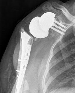
Early closed reduction can be successful. Recurrent or chronic instability may require surgical revision. The case below shows a failed anatomic prosthesis for fracture with anterosuperior escape that was treated with a reverse that dislocated recurrently. 
 

The prosthesis was revised to a hemiarthroplasty that was unsatisfactory. Finally a successful revision was accomplished by the removal of posterior scar tissue and revision to a reverse with a 40 mm set of components.

Shown below is another example where stability was restored by changing to a larger diameter of curvature and increasing the thickness of the polyethylene and humeral cup.

The risk of humeral fracture is increased in revision surgery, by falls and when the humeral component fixation results in an abrupt transition between a cemented or press fit diaphyseal stem tip and osteopenic bone distal to the prosthetic tip These fractures deserve a trial at closed management.  
in that surgical revision can be very complex  

Scapular and acromial fractures can result from excessive deltoid tension producing a fatigue fracture or from bone weakened by screw placement. These fractures are preferably treated non-operatively.

Scapular notching is a prominent complication, particularly common in prosthetic designs that medialize the humerus or when the glenoid component is positioned high on the glenoid bone.


The issues with notching are not so much the ‘notch’ in the scapula but the radiographically unseen damage to the polyethylene of the humeral component
 and to glenoid component fixation 
Notching may also be associated with unwanted bone formation that limits the range of motion 
Humeral component failure may result from dissociation of the cup from the stem Glenoid failure may result from glenosphere-baseplate dissociation, glenoid fracture, or failure of fixation While some cases can be reconstructed, others require salvage conversion to a hemiarthroplasty after glenoid component removal; the clinical results of this conversion are generally poor.

Neurologic lesions can result from dissection, retraction or over lengthening of the arm. Finally, there has been some concern about loss of active external rotation with reverse total designs that medialize the tuberosity, prompting consideration of latissimus dorsi transfers; this problem seems less of an issue with those designs that maintain ‘East-West” tensioning of the residual cuff posteriorly.

domingo, 24 de mayo de 2015

Pectoralis Muscle Anatomy & Function

Fuente
Este artículo es originalmente publicado en:

Educational video describing the muscle anatomy and function of the Pectoralis Muscles.

Become a friend on facebook:
http://www.facebook.com/drebraheim

Follow me on twitter:
https://twitter.com/#!/DrEbraheim_UTMC
  • Categoría

  • Licencia

    • Licencia estándar de YouTube


viernes, 15 de mayo de 2015

Reconstrucción Mínimamente invasiva Tipo IV aguda y Separacion Tipo V acromioclavicular / Minimally Invasive Reconstruction of Acute Type IV and Type V Acromioclavicular Separations


Fuente
Este artículo es originalmente publicado en:
http://www.ncbi.nlm.nih.gov/pubmed/25901627
http://www.healio.com/orthopedics/journals/ortho/2015-4-38-4/%7Bc905d521-004a-4066-a11d-3efdc3ac6527%7D/minimally-invasive-reconstruction-of-acute-type-iv-and-type-v-acromioclavicular-separations#?ecp=318F9B42-3E81-E311-ADF0-A4BADB296AA8
De:
Katsenis DLStamoulis DBegkas DTsamados S.
 2015 Apr 1;38(4):e324-30. doi: 10.3928/01477447-20150402-62.
Todos los derechos reservados para:
Copyright 2015, SLACK Incorporated.


Abstract


The goal of this study was to evaluate the midterm radiologic, clinical, and functional results of the early reconstruction of the severe acromioclavicular joint dislocation using the flipptack fixation button technique. Between December 2006 and December 2009, one hundred thirty-five consecutive patients with acromioclavicular joint separations were admitted to the authors' institution. Fifty patients were included in the study. According to Rockwood classification, 29 (58%) dislocations were type IV and 21 (42%) were type V. Surgery was performed at an average of 4.2 days (range, 0-12 days) after dislocation. All dislocations were treated with the flipptack fixation button technique. All patients were evaluated at a final postoperative follow-up of 42 months (range, 36-49 months). The clinical outcome was assessed using the Constant score. The functional limitation was assessed using the bother index of the short Musculoskeletal Function Assessment. Radiographs taken immediately postoperatively and at the final follow-up assessed acromioclavicular joint reduction, coracoclavicular distance, and joint arthrosis. At the final follow-up, mean Constant score was 93.04 (range, 84-100). The average (±SD) short Musculoskeletal Function Assessment bother index was 20.88±8.95 (range, 2.0-49). No statistically significant difference was found between the acromioclavicular joint dislocation type and the clinical result (P=.227; chi-square, 6.910, Kruskal Wallis test). The regression of the coracoclavicular distance at final follow-up was not statistically significant (P=.276; chi-square, 6.319, Kruskal Wallis test). The flipptack fixation button technique is an effective alternative for the treatment of severe acromioclavicular joint dislocation. Because all objectives of the treatment were obtained, the results do not deteriorate over time. [Orthopedics. 2015; 38(4):e324-e330.].

Resumen
El objetivo de este estudio fue evaluar los resultados radiológicos de mitad de período, clínicos y funcionales de la temprana reconstrucción de la severa 
 luxación acromioclavicular de la articulación mediante la técnica botón de fijación flipptack. Entre diciembre de 2006 y diciembre de 2009, ciento Treinta y cinco pacientes consecutivos con separaciones conjuntas acromioclaviculares  fueron admitidos en la institución de los autores. Cincuenta pacientes se incluyeron en el estudio. Según la clasificación Rockwood, 29 (58%) fueron dislocaciones de tipo IV y 21 (42%) fueron de tipo V. La cirugía se realizó en un promedio de 4,2 días (rango, 0-12 días) después de la dislocación. Todas las dislocaciones fueron tratados con la técnica botón de fijación flipptack. Todos los pacientes fueron evaluados en una final de seguimiento postoperatorio de 42 meses (rango, 36-49 meses). La evolución clínica se evaluó mediante la puntuación de Constant. La limitación funcional se evaluó mediante el índice de molestia de corto Evaluación función musculoesquelética. Las radiografías tomadas inmediatamente después de la operación y al final del seguimiento evaluado acromioclavicular reducción conjunta, distancia coracoclavicular y artrosis articular. Al final del seguimiento, la puntuación media constante era 93,04 (rango, 84-100). La media (± DE) de corto Evaluación Función musculoesqueléticos molesta índice fue de 20,88 ± 8,95 (rango, 2,0-49). No se encontraron diferencias estadísticamente significativas entre el tipo de luxación de la articulación acromioclavicular y el resultado clínico (p = 0,227; chi-cuadrado, 6.910, Kruskal Wallis test). La regresión de la distancia coracoclavicular al final del seguimiento no fue estadísticamente significativa (p = 0,276; chi-cuadrado, 6.319, Kruskal Wallis test). La técnica botón de fijación flipptack es una alternativa eficaz para el tratamiento de la luxación de la articulación acromioclavicular severa. Debido a que se obtuvieron todos los objetivos del tratamiento, los resultados no se deterioran con el tiempo
Copyright 2015, SLACK Incorporated.
PMID:
 
25901627
 
[PubMed - in process]

jueves, 14 de mayo de 2015

Revisión test para #impingements de #hombro y lesiones asociadas de bursa, tendón o labrum

 #impingements  #hombro #shoulder #pinzamiento

Fuente
Este artículo es originalmente publicado en:
http://www.ncbi.nlm.nih.gov/pubmed/23633343
http://onlinelibrary.wiley.com/doi/10.1002/14651858.CD007427.pub2/abstract;jsessionid=6E46497074CA1E349114ED9D3E70140A.f03t04
De:
Hanchard NC1Lenza MHandoll HHTakwoingi Y.
 2013 Apr 30;4:CD007427. doi: 10.1002/14651858.CD007427.pub2.
Todos los derechos reservados para:
Copyright © 1999–2015 John Wiley & Sons, Inc. All Rights Reserved


lunes, 11 de mayo de 2015

I-4 Efecto de las pulsaciones de ultrasonido de baja intensidad (LIPUS) en la expresión de Sclerostin en el proceso de osificación endocondral / I-4 Effect of Low-Intensity Pulsed Ultrasound (LIPUS) on Expression of Sclerostin in the Process of Endochondral Ossification

#LIPUS #osificacion endocondral #sclerostin

Fuente
Este artículo es originalmente publicado en:
http://www.ncbi.nlm.nih.gov/pubmed/25900755
http://journals.lww.com/jorthotrauma/Abstract/2015/05000/I_4_Effect_of_Low_Intensity_Pulsed_Ultrasound.18.aspx
De:
Kumagai K1, Akamatsu Y, Kobayashi H, Kusayama Y, Mitsuhashi S, Kobayashi A, Saito T.
J Orthop Trauma. 2015 May;29(5):S3. doi: 10.1097/01.bot.0000462955.71988.6d.
Todos los derechos reservados para:
Copyright © 2015 Wolters Kluwer Health, Inc. All rights reserved.


Abstract

OBJECTIVE:

We have previously demonstrated that LIPUS inhibited expression of sclerostin and stimulated new bone formation in a mouse osteotomy model. To further address effect of LIPUS on endochondral ossification, expression of sclerostin was examined in vitro.
RESULTS:Sclerostin was identified in the culture media at 3 and 5 weeks, and the concentration in LIPUS group was significantly less than that in the control group. PCR analysis demonstrated that sclerostin gene expression was significantly decreased in the LIPUS group in comparison to the control group at 3 and 5 weeks. And gene expressions of Col2a1 and Col10a1 were significantly decreased in the LIPUS group at 5 weeks.
CONCLUSIONS:This study suggested that expression of sclerostin in the process of endochondral ossification was inhibited by LIPUS treatment.

Resumen

OBJETIVO:
Hemos demostrado previamente que LIPUS inhibió la expresión de esclerostina y estimuló la formación de hueso nuevo en un modelo de ratón de la osteotomía. Para mayor efecto dirección del LIPUS en la osificación endocondral, la expresión de esclerostina fue examinada in vitro.
RESULTADOS:
La esclerostina fue identificada en los medios de cultivo a los 3 y 5 semanas, y la concentración en el grupo LIPUS fue significativamente menor que en el grupo control. El análisis por PCR demostró que la expresión de genes esclerostina se redujo significativamente en el grupo LIPUS en comparación con el grupo control a los 3 y 5 semanas. Y las expresiones de genes de Col2a1 y Col10a1 se redujeron significativamente en el grupo LIPUS a las 5 semanas.
CONCLUSIONES:
Este estudio sugiere que la expresión de esclerostina en el proceso de osificación endocondral fue inhibida por el tratamiento LIPUS.
PMID:
 
25900755
 
[PubMed - in process]

sábado, 9 de mayo de 2015

Fracturas del húmero distal.

Fuente
Este artículo es originalmente publicado en:
http://www.slideshare.net/JenaroMD/fracturas-del-hmero-distal-curso-de-instruccin-sociedad-espaola-ciruga-hombro-y-codo

(4 SlideShares) , Especialista Senior en Cirugía Ortopédica y Traumatología. Unidad de Cadera. at Unidad de Cadera. Servicio COT. Hospital Clínic Barcelona (Spain)